Hinde Lab: Cellular Biophysics
The Laboratory for Cellular Biophysics develops optical microscopy methods based on fluorescence lifetime, anisotropy and correlation spectroscopy to spatiotemporally map the cell nucleus and uncover the role of nuclear architecture in genome function.
Our research focuses on the cell nucleus, where genome function is governed by the dynamic interplay between nuclear architecture and protein navigation. To study these processes in living cells, we develop methods based on fluorescence lifetime and anisotropy imaging microscopy (FLIM and FAIM) of Förster resonance energy transfer (FRET), coupled with fluorescence correlation spectroscopy (FCS). These methods allow us to bypass the diffraction limit of optical microscopy and quantify chromatin structure at the nanoscale while tracking protein diffusion with single-molecule resolution.
University of Melbourne
Phone
13 MELB (13 6352)
International
+61 3 9035 5511
Location
David Caro Building
Grattan Street, Parkville
Victoria 3010, Australia
Contact
For enquiries, please email A/Prof Elizabeth Hinde - elizabeth.hinde@unimelb.edu.au
Architectural organisation of the cell nucleus
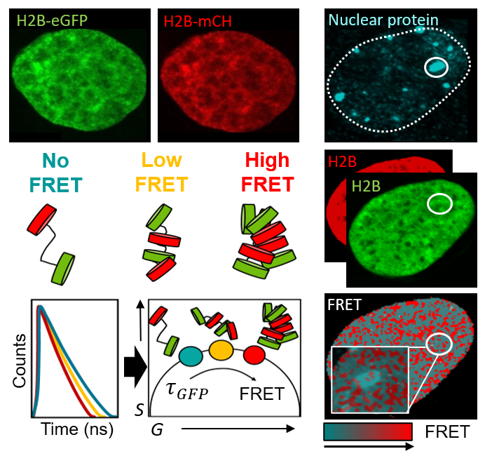
We aim to understand how the two metres of DNA that underpin the human genome are packaged into a 3D chromatin network that fits within the tiny volume of a cell nucleus, and how sub-micron to nanoscale rearrangements in this framework regulate access to the DNA template. To investigate this, we: (1) spatiotemporally map chromatin accessibility via fluorescence fluctuation spectroscopy (FFS) of inert fluorescent tracers of different sizes, and (2) quantify FRET interactions between fluorescently labelled histones (H2B) and architectural proteins (HP1) using FLIM and FAIM. This allows us to study the role of chromatin structure and dynamics in live-cell genome function down to the level of nucleosome proximity.
Relevant publications
- Lou J., Deng Q., Zhang X., et al. Heterochromatin protein 1 alpha (HP1α) undergoes a monomer to dimer transition that opens and compacts live cell genome architecture. Nucleic Acids Res. (2024)
- Solano A., Lou J., Scipioni L., et al. Radial pair correlation of molecular brightness fluctuations maps protein diffusion as a function of oligomeric state within live-cell nuclear architecture. Biophysical Journal (2022)
- Lou J., Scipioni L., Wright B.K. & Hinde E. Phasor histone FLIM-FRET microscopy quantifies spatiotemporal rearrangement of chromatin architecture during the DNA damage response. PNAS USA (2019)
Protein navigation of the nuclear landscape
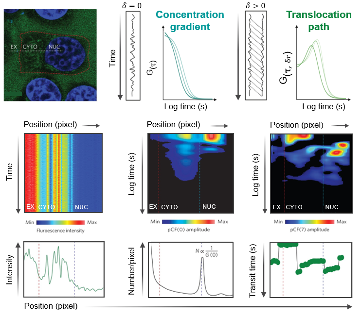
We investigate how DNA-binding proteins navigate the nucleus and how protein–protein interactions regulate DNA target search. To do so, we are developing fluorescence fluctuation-based analyses to extract nuclear trafficking events directly from live-cell microscopy data. Using spatiotemporal correlation functions, we map protein local diffusion to long-range nuclear transport, while resolving how these dynamics shift with oligomeric state or complex formation via use of spectrally distinct fluorescent ligands. These measurements are compatible with single-, dual-, or triple-channel CLSM or TIRF microscopy time series and allow us to determine how DNA repair factors navigate nuclear architecture to coordinate double-strand break resolution, and how transcription factors use protein stoichiometry to regulate DNA target search and gene expression.
Relevant publications
- Hinde E., Thammasiraphop K., Duong H. et al. Pair correlation microscopy reveals the role of nanoparticle shape in intracellular transport and site of drug release. Nature Nanotech. (2017)
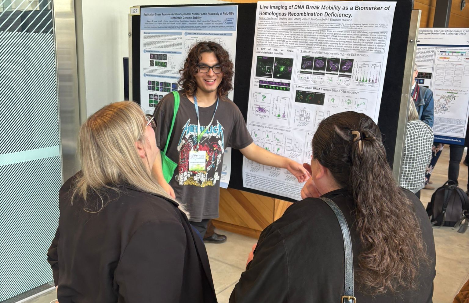
- Lou J., Priest D.G., Solano A., et al. Spatiotemporal dynamics of 53BP1 dimer recruitment to a DNA double strand break. Nat Commun. (2020)
- Sanchez-Velasquez J., Solano A., Digman M.A., et al Pair correlation microscopy of intracellular molecular transport. Nat Protoc. (2025)
Advances in optical microscopy and molecule-specific fluorescence labelling strategies have enabled molecular insights into the spatiotemporal dynamics of biological processes that were previously inaccessible.
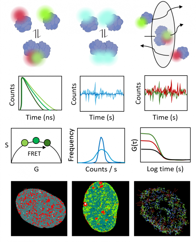
In the Laboratory for Cellular Biophysics, we specialise in parallel measurement of multiple fluorescence observables – intensity, anisotropy, and lifetime – to quantify molecular behaviour in the native environment of the living nucleus. By coupling these observables with FRET and fluorescence correlation spectroscopy (FCS), we can resolve: (1) structure at the nanometre scale, using FRET to detect protein–protein and protein–ligand interactions, and (2) dynamics across nanoseconds to milliseconds, using FCS and related approaches to quantify diffusion, transport, and interaction kinetics. This methodological framework enables us to measure key molecular parameters, including protein–ligand binding, oligomerisation, and intracellular trafficking.
Core methods
FLIM Fluorescence lifetime imaging microscopy: used for hetero-FRET detection and mapping of protein interactions, as well as biosensing of intracellular pH.
FAIM Fluorescence anisotropy imaging microscopy: used for homo-FRET detection and mapping of protein self-association, as well as biosensing of intracellular viscosity.
FCS Fluorescence correlation spectroscopy: used to quantify local protein dynamics in a subcellular location (e.g. diffusion coefficient and concentration).
FFS Fluorescence fluctuation spectroscopy and brightness analysis: used to quantify and spatially map protein oligomer formation and stoichiometry.
sFCS, pCF Scanning FCS and pair correlation function analysis: used to quantify protein transport with respect to subcellular architecture (e.g. arrival time, directionality, diffusion law).
STICS, RICS Spatiotemporal image correlation spectroscopy: used to map protein mobility and binding dynamics across a subcellular location.
Together, these methods provide a powerful framework to reveal how molecular interactions and dynamics underpin genome function within the context of live-cell nuclear architecture.
Equipment available
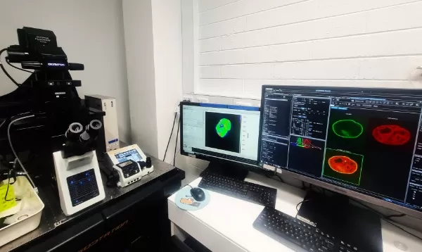
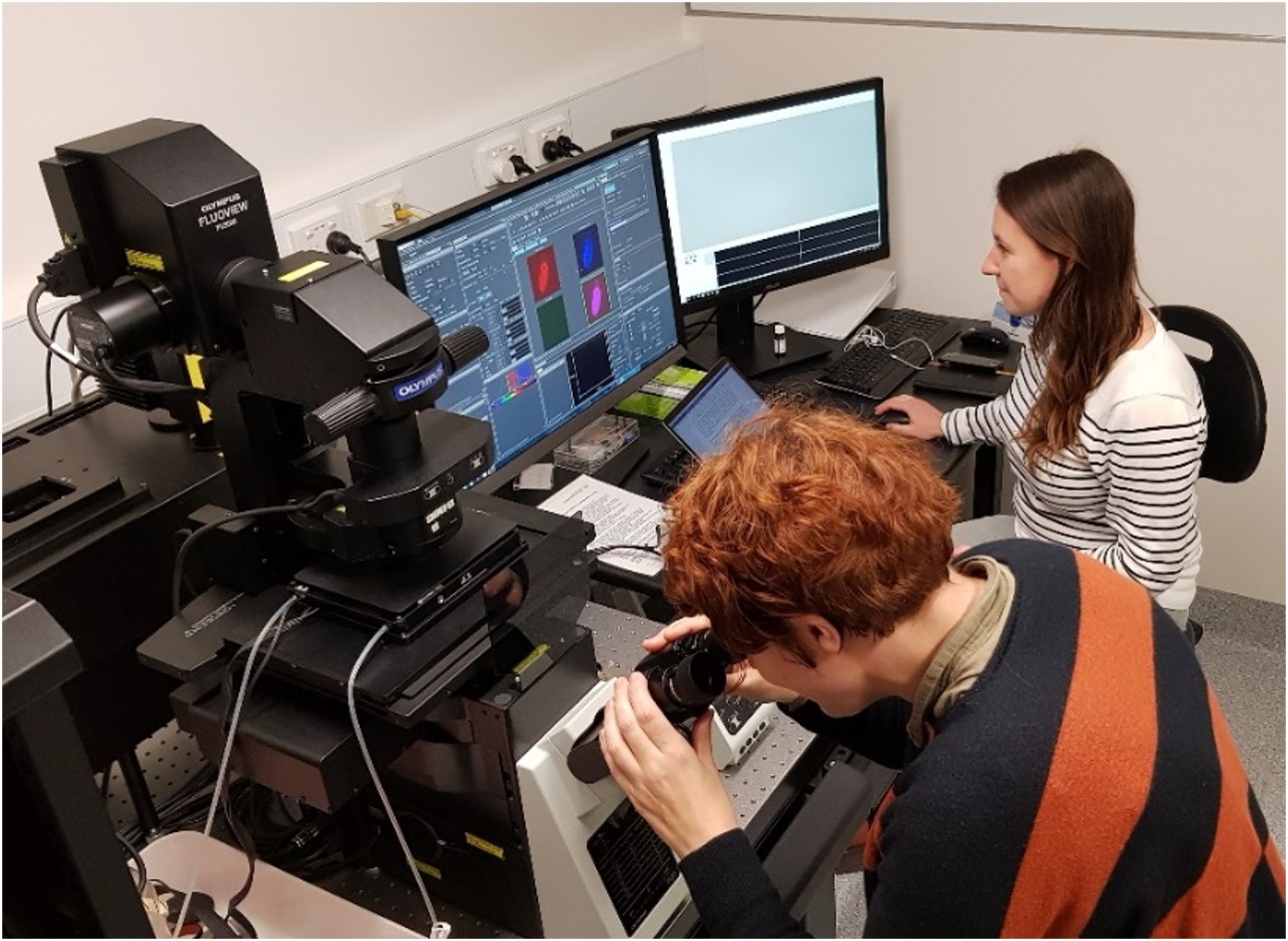
Olympus FV3000
High-performance confocal laser scanning microscope coupled to an ISS FLIMbox, enabling time-resolved fluorescence lifetime detection alongside fluorescence fluctuation spectroscopy and orbital tracking for quantitative live-cell imaging.
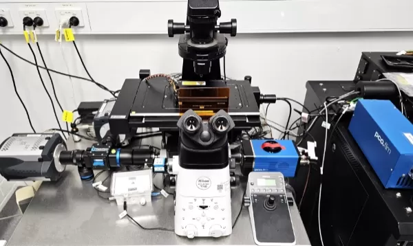
Nikon Ti2 Eclipse
High-performance TIRF and widefield fluorescence microscope coupled to a time-resolved PCO camera and pulsed laser excitation, enabling lifetime-based FRET detection and single-molecule tracking for quantitative live-cell imaging.
Learn more about our equipment
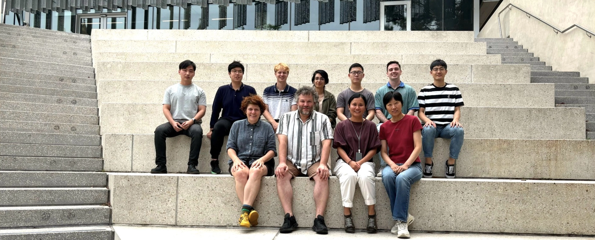
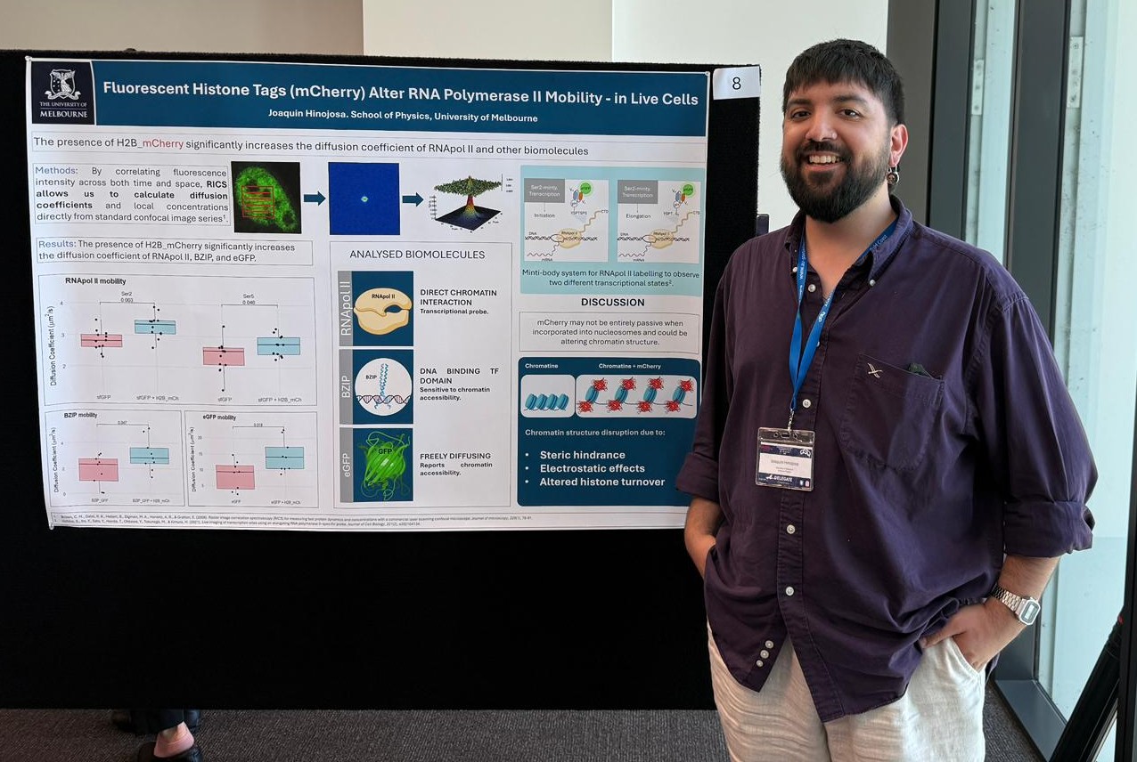
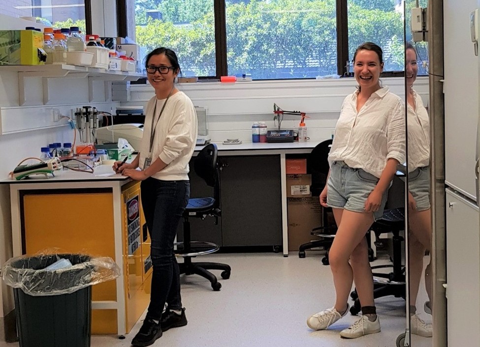
Lab members


Meet us
Alumni
- Saul Menendez Cardenas (2025 Honours student)
- Siyuan Meng (2025 Master student)
- Paul Armitage (2025 Exchange student)
- Blake Bishop (2025 Master Student)
- Tao Sun (2024 Master student) now PhD student at UoM
- Ashleigh Solano (2024 PhD student) now Post-doc at WEHI
- Dominic Gehrig (2024 Honours student)
- Yifu Zheng (2023 Master student)
- Kedi Wang (2023 Master student)
- Niall Karunaratne (2023 Exchange student)
- Alex Hopper (2023 Master student)
- David Priest (2023 Post-doc)
- Adele Kerjouan (2023 Post-doc)
- Zhen Liang (2022 Master student)
Selected publications
For a complete list of publications
2025
Sanchez-Velasquez J., Solano A., Digman M.A., Gratton E., Cardarelli F. & Hinde E. Pair correlation microscopy of intracellular molecular transport. Nat Protoc. 2025. 10: 1651–1677. DOI: 10.1038/s41596-024-01097-6
2024
Lou J., Deng Q., Zhang, X., Bell C.C., Das A.B., Bediaga N.G., Zlatic C.O., Johanson T.M., Allan R.S., Griffin M.D.W., Paradkar P., Harvey K.F., Dawson M.A. & Hinde E. Heterochromatin protein 1 alpha (HP1α) undergoes a monomer to dimer transition that opens and compacts live cell genome architecture. Nucleic Acids Research 2024. 52(18): 10918-10933. DOI: 10.1093/nar/gkae720
Lian Z., Solano A., Lou J. & Hinde E. Histone FRET reports the spatial heterogeneity in nanoscale chromatin architecture that is imparted by the epigenetic landscape at the level of single foci in an intact cell nucleus. Chromosoma 2024. 133(1): 5-14. DOI: 10.1007/s00412-024-00815-z
Mehl B.P., Vairaprakash P., Li L., Hinde E., MacNevin C.J., Hsu C., Gratton E., Liu B. & Hahn K.M. Live-cell biosensors based on the fluorescence lifetime of environment-sensing dyes. Cell Reports Methods 2024. 4(3): 100734. DOI: 10.1016/j.crmeth.2024.100734
2023
Owyong T.C., O’Shea R., Lee M., White J.M., Donnelly P.S., Hinde E., Wong W.W.H. & Hong Y. A General Fluorescence-Based Method for Quantifying and Mapping Biomolecular Polarity In Vitro and In Cells. bioRxiv 2023. DOI: 10.1101/2023.02.07.526546
2022
Solano A., Lou J., Scipioni L., Gratton E. & Hinde E.. Radial pair correlation of molecular brightness fluctuations maps protein diffusion as a function of oligomeric state within live-cell nuclear architecture. Biophysical Journal 2022. 122(11): 2152-2167. DOI: 10.1016/j.bpj.2022.04.030
Mitrentsi, I., Lou J., Kerjouan A., Verigos J., Reina-San-Martin B., Hinde E. & Soutoglou E. Heterochromatic repeat clustering imposes a physical barrier on homologous recombination to prevent chromosomal translocations. Mol Cell. 2022. 82(11): 2132–2147.e6 DOI: 10.1016/j.molcel.2022.03.033
Deore P., Wanigasuriya I., Tsang Min Ching S.J., Brumley D.R., van Oppen M.J.H., Blackall L.L. & Hinde E.. Fluorescence lifetime imaging microscopy (FLIM): a non-traditional approach to study host-microbial symbioses. Micriobiol Aust. 2022. 43(1): 22-27 DOI: 10.1071/MA22008
2021
Lou J., Solano A., Liang Z. & Hinde E.. Phasor Histone FLIM-FRET Microscopy Maps Nuclear-Wide Nanoscale Chromatin Architecture With Respect to Genetically Induced DNA Double-Strand Breaks. Front Genet. 2021. 12: 770081. DOI: 10.3389/fgene.2021.770081
Priest D.J., Bernardini A., Lou J., Mantovani R. & Hinde E.. Live cell dynamics of the NF-Y transcription factor. Sci Rep. 2021. 11: 10992 DOI: 10.1038/s41598-021-90081-1
Wesemann L., Rickett J., Song J., Lou J., Hinde E., Davis T.J. & Roberts A. Nanophotonics enhanced coverslip for phase imaging in biology. Light Sci Appl. 2021. 10: 98 DOI: 10.1038/s41377-021-00540-7
Palmer C.S., Lou J., Kouskousis B., Pandzic E., Anderson A.J., Kang Y., Hinde E. & Stojanovski D. Super-resolution microscopy reveals the arrangement of inner membrane protein complexes in mammalian mitochondria. J Cell Sci. 2021. 134(13): jcs252197. DOI: 10.1242/jcs.252197
2020
Lou J., Priest D.G., Solano A., Kerjouan A. & Hinde E.. Spatiotemporal dynamics of 53BP1 dimer recruitment to a DNA double strand break. Nat Commun. 2020. 11(1): 5776. DOI: 10.1038/s41467-020-19504-3
Liang Z., Lou J., Scipioni L., Gratton E. & Hinde E.. Quantifying nuclear wide chromatin compaction by phasor analysis of histone Förster resonance energy transfer (FRET) in frequency domain fluorescence lifetime imaging microscopy (FLIM) data. Data Brief 2020. 30: 105401. DOI: 10.1016/j.dib.2020.105401
2019
Priest D.J., Solano A., Lou J. & Hinde E.. Fluorescence fluctuation spectroscopy: an invaluable microscopy tool for uncovering the biophysical rules for navigating the nuclear landscape. Biochem Soc Trans. 2019. 47(4):1117-1129. DOI: 10.1042/BST20180604
Lou J.,Khor J., Priest D. & Hinde E.. Fluorescence Fluctuation Spectroscopy Reveals Double Strand Break Recruitment of 53BP1 Dimers and Assembly into Higher-Order Oligomers at the DNA Repair Locus. Biophysical Journal. 2019. 116(3) DOI: 10.1016/j.bpj.2018.11.156
Lou J., Scipioni L., Wright B.K. & Hinde E.. Phasor histone FLIM-FRET microscopy quantifies spatiotemporal rearrangement of chromatin architecture during the DNA damage response. Proceedings of the National Academy of Sciences. 2019. 116(15): 7323-7332 DOI: 10.1073/pnas.1814965116
2017
Hinde E., Thammasiraphop K., Duong H.T.T., Yeow J., Karagoz B., Boyer C., Gooding J. & Gaus K. Pair correlation microscopy reveals the role of nanoparticle shape in intracellular transport and site of drug release. Nature Nanotech. 2017. 12: 81–89. DOI: 10.1038/nnano.2016.160
2016
Hinde E., Pandžić E., Yang Z., Ng I.H.W., Jans D.A., Bogoyevitch M.A., Gratton E. & Gaus K. Quantifying the dynamics of the oligomeric transcription factor STAT3 by pair correlation of molecular brightness. Nat Commun. 2016. 7: 11047. DOI: 10.1038/ncomms11047
2015-2014
Hinde E., Cardarelli F. & Gratton E. Spatiotemporal regulation of Heterochromatin Protein 1-alpha oligomerization and dynamics in live cells. Sci Rep. 2015. 5: 12001. DOI: 10.1038/srep12001
Hinde E., Kong X., Yokomori K. & Gratton E. Chromatin dynamics during DNA repair revealed by pair correlation analysis of molecular flow in the nucleus. Biophys J. 2014. 107(1): 55-65. DOI: 10.1016/j.bpj.2014.05.027